Restoration in progress

Mary Turner nearly fainted when her doctor told her she had carcinoma in her right eye. “It just floored me,” she says.
Treating the cancer required surgery to remove her eye and surrounding tissues. The desire to restore her appearance through a prosthesis anchored to craniofacial implants led to her referral to the Center for Maxillofacial Prosthodontics at Texas A&M University Baylor College of Dentistry.
By the time she completed her care, her silicone prosthesis was hardly detectable during an on-camera interview by the news crew of Univision Dallas, a Spanish-language television station that covered the center’s work in a two-part series.
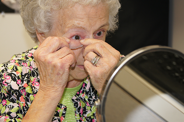
“Oh my goodness,” Turner exclaimed when she first saw her prosthesis in place. She looked up from the hand mirror and beamed at her son and the gathered faculty and staff.
The center, one of just a few multidisciplinary ones in the U.S. and Canada that include an anaplastologist – a specialist in restoring a malformed or missing part of the human body through artificial means – is the only one of its type housed within a dental school.
Ongoing collaboration with faculty members of TAMBCD’s Department of Oral and Maxillofacial Surgery, including Turner’s implant surgeon Dr. Marianela Gonzalez, assistant professor, promotes advanced presurgical planning and navigational surgery: cutting-edge techniques transforming care.
“It is imperative to have a thorough preoperative plan to make sure the craniofacial implants are placed into an area with good bone quantity, and in a location that will provide a good prosthetic result,” says Suzanne Verma, assistant professor and anaplastologist. “We precisely plan the implant locations for patients based on their CT scans, enabling us to go straight into the operating room with the digital plan.
“Our surgeries use navigational technology, otherwise explained as ‘GPS in the OR.’ When our instruments touch a patient in surgery, we can see where we are in the CT scan and our digital plan in real time.”
Verma can manipulate the same radiographic data to create a physical model of the missing anatomy.
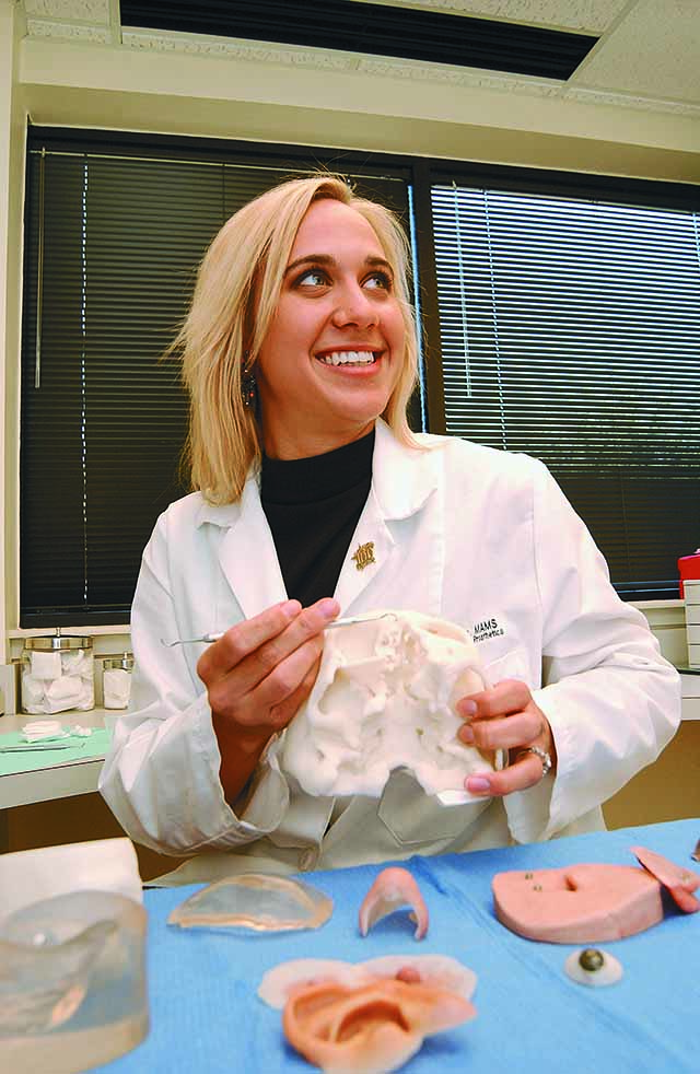
Four to six months following surgery, after the implants are fully integrated with the bone and the soft tissue is healed, the patient returns for a series of appointments with Verma, who uses a combination of art, science and digital technology to create a prototype and mold for the patient’s silicone prosthesis.
She still spends up to a week in the clinic and lab – longer for more complex cases – creating the perfect prosthesis by pigmenting silicone to perfectly match the patient’s skin tone, vascularization and unique characteristics. This process often requires as many as 16 different colors painted into the mold layer by layer.
The scope of technological advancements in Verma’s field is far-reaching. She is involved on an international level with a multidisciplinary group called Advanced Digital Technology in Head and Neck Reconstruction, which she serves as a member of its scientific advisory board.
“We meet every three years, and it’s amazing to hear how interdisciplinary craniofacial teams around the world are applying new technology to patient care,” Verma says.
In addition to department faculty, Verma collaborates on navigational surgery with medical and dental professionals at numerous hospitals in North Texas and receives referrals from around the U.S. and South America.
Media contact: media@tamu.edu


